This video describes the basic approach to interpret and report a trauma radiograph X-Ray filmNEET PG EntranceFracture X rayFor more topics in Orthopaedi. On your exam keep an eye out for small poke holes in skin.
2
A systematic approach to describing fractures JAAPA.

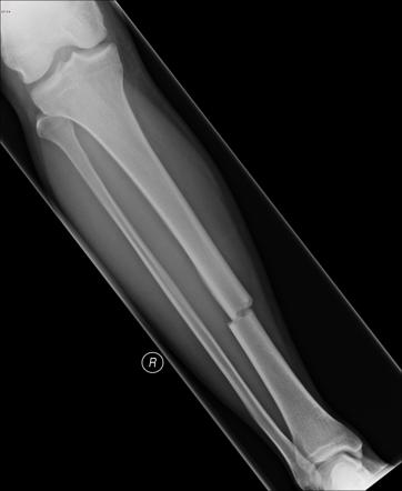
. Introduction to Trauma X-ray Other fracture types Key points An avulsion injury arises when a bone fragment is pulled away by a ligament or tendon A stress fracture arises in a normal bone as a result of repetitive low impact trauma Periprosthetic fractures occur. The radiograph below demonstrates why. The number of fragments.
Study How to Describe a Fracture X-rays flashcards from Priyanka Bhandaris University of Aberdeen class online or in Brainscapes iPhone or Android app. Non-union pseudoarthrosis fracture healing does not occur within 6-9 months. In most instances the fracture is evident clinically and easily identified on radiographs.
Keaton Jones defines a fracture and shows you an easy method to describe any fracture with examples from x-rays of patients. Fractures are usually described using four major parameters Table 22-3. A fracture with an offset of 2 mm or more in any plane or 2 mm offset involving the articular surface is considered displaced.
Transverse oblique spiral Simple or. Details of the radiograph and the patient. Normal fracture healing can be disrupted in numerous ways.
When describing the displacement the position of the fragment distal to the fracture site is always described. Disturbed fracture healing. Introduction to Trauma X-ray Fracture description Key points Fracture description depends on the class of bone and the direction of the fracture line A long bone fracture is described according to its direction in relation to the shaft of the bone Bones and fractures There are 4 anatomical classes of bone - long short flat and irregular.
X-ray appearances of fracture complications and some common fracture mimics are also described. Annelies van der Plas MSK radiologist Maastricht UMC. Diastasis example - Pelvis Hover onoff image to showhide findings Click image to align with top of page Diastasis example - Pelvis.
Start studying X-ray describing fractures. There is a common lexicon used in describing fractures to facilitate a reproducible description and to assure reliable and accurate communication. Axial shortening radial inclination and radio-ulnar displacement can be measured on the routine posterioranterior film.
First Steps Ensure that you are looking at the correct patient - check the name and date of birth. Learn vocabulary terms and more with flashcards games and other study tools. Non-displaced fractures may be subtle on x-ray Report checklist AP pelvis and lateral hip should be viewed because pelvic fractures can mimic clinical features of hip fracture trace Shentons line assess for symmetry particularly note lesser trochanter may indicate external rotation bone trabeculae sclerosis smudge Treatment and prognosis.
The direction of the fracture line. Complete transverse oblique spiral comminuted incomplete buckle greenstick fracture location. It is common for clavicle fractures to be displaced due to a combination of the weight of the upper limb pulling the distal fragment down and the sternocleidomastoid pulling the medial fragment upwards.
Characteristics of the fracture such as the type deformity and soft tissue joint involvement are used to guide management. Part of the bone epiphysis metaphysis diaphysis apophysis some bones have specific named parts eg. Introduction to Trauma X-ray Fracture displacement Key points Describe fracture displacement in terms of the abnormal position of the distal fragment in relation to the proximal bone Displacement of fractures is defined in terms of the abnormal position of the distal fracture fragment in relation to the proximal bone.
Radiologic reporting of skeletal traumaM J MJ Pitt and D P DP Speer Radiol Clin North Am 28 2247-56 1990 BJ. Scaphoid bone femoral neck tibial shaft. Author Sarah Bolander 1 Affiliation 1 Sarah Bolander is an assistant professor at Midwestern University in Glendale Ariz and practices at Cactus Pediatric Orthopedics in Mesa Ariz.
This video podcast is essential. For 25- you have access to all modules. Fracture healing takes about twice as long as expected for a specific location.
The common terminology used for describing fractures is discussed. Bleeding from bone is often constant slow venous type bleeding. Basic concepts are introduced regarding the use of X-rays in management of fractures and dislocations.
Which bone which part of the bone left or right. Changes in alignment suggest a fracture subluxation partial dislocation or dislocation. First thing to mention when describing a fracture.
You cant always tell if a fracture is open or closed from the x-rays always use orthogonal views xrays that are 90 degrees from each other such as an AP and lateral. Displacement can be dorsal volar radial or proximal. Describing a fracture on an ankle radiograph.
Type of fracture eg. When describing an ankle X-ray use the following structure. Click for a list of eponymous fractures X-rays are commonly used in clinical practice to diagnose fractures.
The combined injuries are termed fracture-dislocation Diastasis Diastasis is a term used to describe the separation of 2 normally adjacent bone parts either at a ligamentous joint or at a growth plate. The author has disclosed no potential. 24012014 translated 23082016 Sources.

Describing A Fracture An Approach Radiology Reference Article Radiopaedia Org

Define The Parameters That Allow You To Describe A Long Bone Fracture Pattern Over The Phone Orthopaedia
Describing A Fracture An Approach Radiology Reference Article Radiopaedia Org
Describing A Fracture An Approach Radiology Reference Article Radiopaedia Org


0 Comments